Clinical Imaging Engine
live Domain: clinical Type: engine
Live DICOM analysis on real pixels: the live-verified modality is chest X-ray via DenseNet-121, with FHIR R4 export and cross-modality reasoning against genomics. The CT path (VISTA-3D segmentation, VILA-M3 image/report reasoning) is planned/placeholder, not yet live. MAISI synthetic imaging is research/augmentation/QA only, never diagnostic. Decision support. The Cardiac Workup (CT coronary angiography) workflow is REPRESENTATIVE: ct_coronary_angiography.infer() returns fixed findings (CAD-RADS 4A/P3/HRP, Agatston 385 representative, 77.9% stenosis measured from a segmented vessel surface) - SegResNet/VISTA-3D coronary inference is not yet wired. Its imagery is a rendered coronary surface mesh (CoronariesNC6, manual rater ground truth), not a patient CT.
A narrated, captioned explainer. Decision support for a qualified clinician.
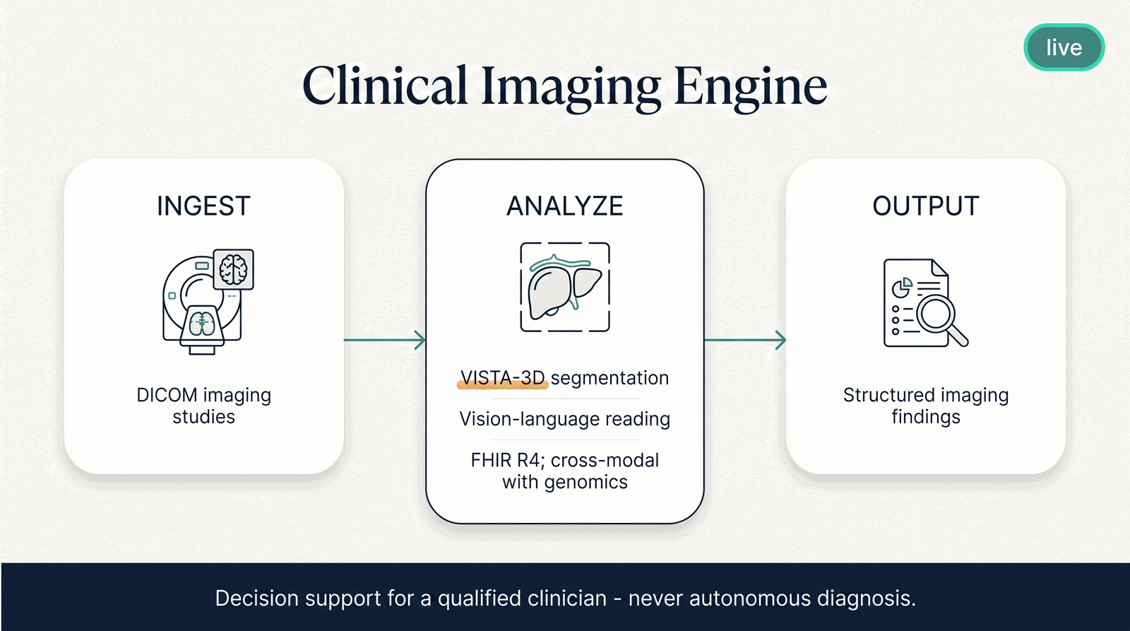
Illustrative. Decision support for a qualified clinician — never autonomous diagnosis or prescribing.
In plain terms
The Clinical Imaging Engine reads medical images and reasons about them alongside the patient's genomics. It turns a scan — delivered as standard DICOM — into structured findings a clinician can act on, and it can cross-reference those findings against what the genome says. Image and gene together often say more than either alone.
Why it matters
A radiology read and a genomic finding usually live in separate systems and separate heads. Bringing them into one reasoning step — a calcium score next to a lipid genotype, a tumor's imaging next to its fusion — is where cross-modal precision medicine actually happens.
For a patient: their scan and their genetics read together, so nothing that matters falls through the gap between two systems.
How it works
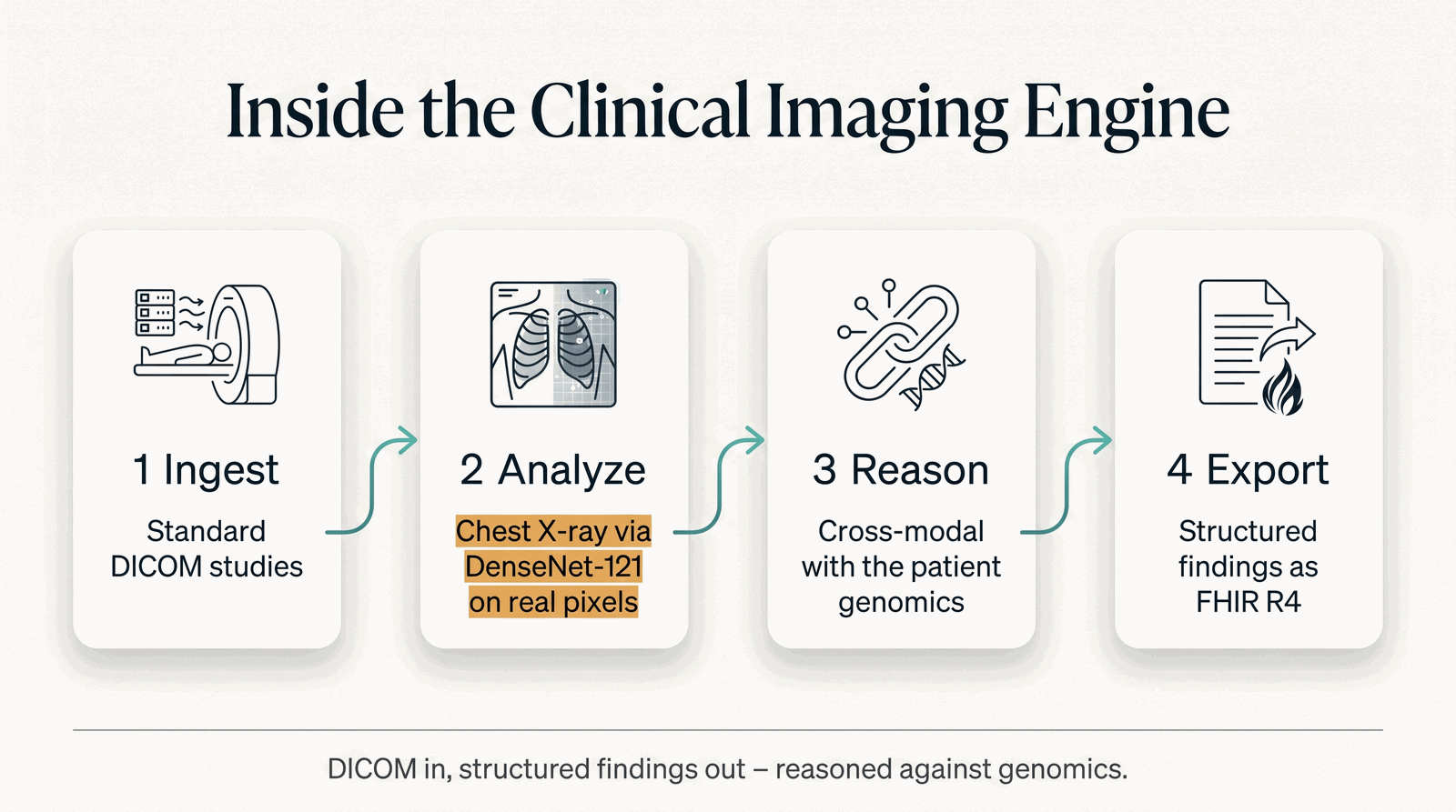
DICOM in, structured findings out — reasoned against genomics. Illustrative.
- Ingest — a standard DICOM imaging study comes in (referenced through the patient context).
- Analyze — the live, verified modality today is chest X-ray analysis via a DenseNet-121 model on real DICOM pixels.
- Reason — that live read is joined cross-modally with the patient's genomics (the deeper image-and-report reasoning, VILA-M3 on CT, is on the planned path — see Honest limits).
- Export — findings are written out as FHIR R4 so they slot into clinical systems.
What goes in, what comes out
- In: a query and the patient context (which references the DICOM study).
- Out: a structured image read.
Where it fits
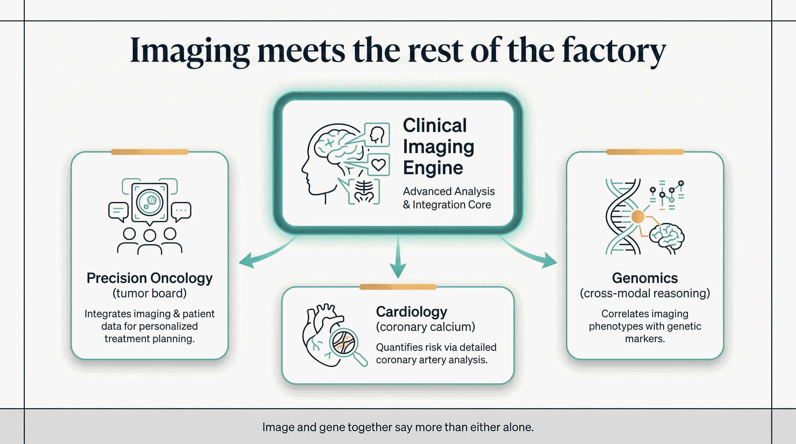
Imaging joins the tumor board, the calcium-score-to-statin path, and genomic reasoning. Illustrative.
Its reads feed the Precision Oncology Engine (imaging joins the tumor board) and the Cardiology Engine (a coronary-calcium input — a CT-derived value, on the planned CT path, not produced by the live chest-X-ray model), and they reason cross-modally with genomics throughout.
Honest limits
- What's live vs. planned. The chest X-ray / DenseNet-121 path is the live-verified modality. The VISTA-3D segmentation and VILA-M3 CT path is planned / placeholder — not yet live.
- Synthetic imaging is not diagnostic. MAISI synthetic image generation is for research / augmentation / QA only — never a diagnostic source.
- Decision support, not diagnosis. The read supports a qualified clinician; it does not diagnose on its own.
Interface
- Endpoint:
localhost:8523· Invoke path:/ - Serving: native · GPU: no · Cost class: —
Inputs
| Name | Shape | Semantic | Notes |
|---|---|---|---|
question |
scalar | — | Field name is question — verified against the live route, which rejects query with 422. |
patient_context |
map | patient_context |
Outputs
| Name | Shape | Semantic | Notes |
|---|---|---|---|
answer |
map | image_read |
Tags: engine · imaging
↩ Back to the Engines index · the Capability Maturity Matrix · the Capability Brief.
Note
Status and interface are generated from the capability registry (lib/hcls_common/capabilities.json) — the site cannot claim ahead of the code. All clinical output is decision support for a qualified clinician, never autonomous diagnosis.